Illustrations by Ron Guastaferri
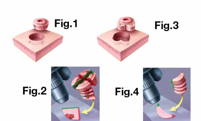
[spacer=’3′]• The site of the cancer is prepared and numbed with local anesthetic. The superficial part that appears to be cancerous, based on color, texture and other factors that distinguish it from the surrounding skin, is removed with a sharp scalpel. (Fig. 1) This piece is marked to determine which side is up, down, etc. The area from where the cancer was removed is cauterized and a temporary dressing is put in place.
• The patient waits while the removed tissue is taken to the lab to prepare. The tissue is separated and the edges are inked with different colors so that the orientation of each piece is preserved. Then, the tissues are frozen in a cryostat—a fast freezer. Once frozen, very thin slices, a few thousandths of a millimeter thick, are removed from the edges of each piece by a sharp blade called a microtome. These are then placed sequentially on glass slides. (Fig. 2) The slides are put through a staining machine where various colors can be used to make the tumor cells stand out from the normal skin cells. Staining is necessary because tissue is otherwise transparent and distinguishing healthy cells from cancer cells would prove too difficult.
• Dr. Alam functions as both the surgeon and pathologist, as it has been shown that this increases the accuracy of the procedure. He looks at the debulk specimen, the center of the lesion, to see the tumor, then he looks at the edges and base of the actual stage that was removed. (Fig. 2) He checks to see if there is any tumor of the type seen in the debulk specimen still present on the outside or bottom edge of the tissue specimen. If microscopic examination shows that the cancer has been completely removed, the patient’s wound is repaired. However, if the doctor sees tumor cells on the edges, he marks them on a map—a physical drawing of the specimen.
• Bearing the map, Dr. Alam returns to the operating site and again prepares the patient for surgery. More skin is removed at just those points along the edges of the prior surgery where the map indicated that some of the tumor remained. (Fig. 3) Again, the wound is cauterized and bandaged and asked to wait while this tissue is processed and reviewed in the lab. These steps are repeated until all of the tumor is removed as evidenced by the tissue showing margins clear of cancer cells. (Fig. 4)


